Tumor suppressor promotes some acute myeloid leukemias, study reveals

3f3ff19d15460279cd9e094bdc487343
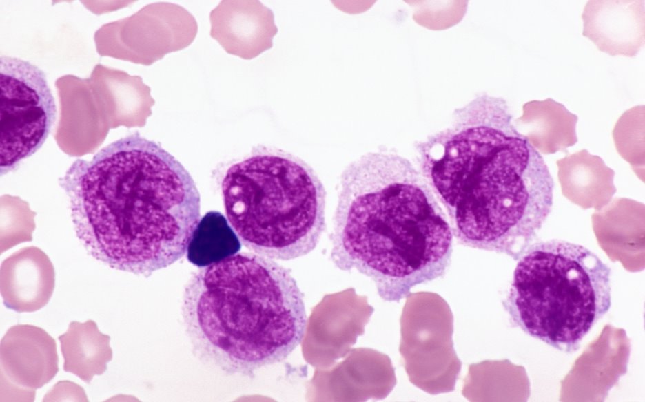
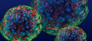
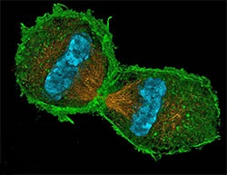
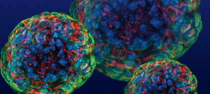
Mutant hematopoietic stem cells in the bone marrow proliferate and fail to differentiate into white blood cells in Acute myeloid leukemia (AML). FLT3 is one of the most common genes mutated; however mutated FLT3 alone is not sufficient in leading to AML. Researchers have found that some patients with mutated FLT3 often have elevated levels of another protein RUNX1, which has been believed to be a tumor suppressor. Surprisingly, they reported that coexpression of RUNX1 with FLT3 blocks the differentiation of white blood cells by inducing another transcription factor Hhex. They proposed RUNX1 may suppress activation of AML initially; however, FLT3 inactivation may lead to RUNX1 activation that would promote tumor growth. Reducing RUNX1 in cancer stem cells may be deleterious but not in normal cells thus providing a selective target for potential interventions.


References:

1.https://www.sciencedaily.com/releases/2017/02/170217095930.htm

2.Kira Behrens, Katrin Maul, Nilgün Tekin, Neele Kriebitzsch, Daniela Indenbirken, Vladimir Prassolov, Ursula Müller, Hubert Serve, Jörg Cammenga, Carol Stocking. RUNX1 cooperates with FLT3-ITD to induce leukemia. The Journal of Experimental Medicine, 2017; jem.20160927 DOI

Article Summary Courtesy: Jennifer Lu and Waleed Khan

Improving longevity of functionally integrated stem cells in regenerative vision therapy

improvinglon
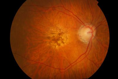
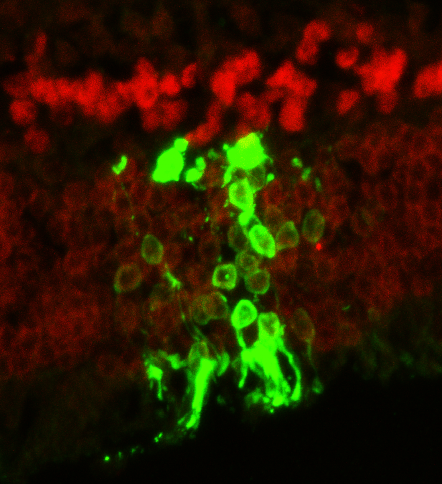
One of the major obstacles in stem cell transplantation is preventing immune rejection that would recognize transplanted cells as foreign and attack them leading to disappointing long-term outcomes. Although the eye and the brain have been considered as exceptions from immune surveillance, Zhu et al. showed that immune rejection was one of the major contributors to degenerated transplanted photoreceptors in blind mice after temporarily restoring sights. Transplantation of photoreceptors derived from stem cells into a strain of immunodeficient IL2 receptor gamma null mice was found to have a 10-fold increase in living transplanted cells that differentiated into mature photoreceptors expressing opsins. Even after nine months to one year after transplantation, the eyes of the mice were shown to be functional and transmitting signals to the brain. Their results showed that to improve longevity of transplanted retinal cells in restoring eye sight, immunosuppression is crucial and specific inhibitors against the IL2 gamma receptor could be developed to selectively suppress immune rejection to retinal cells instead of systemic immunosuppression.

References:

1.https://www.sciencedaily.com/releases/2017/01/170112141243.htm

2.Jie Zhu et al. Immunosuppression via loss of IL2rγ enhances long-term functional integration of hESC-derived photoreceptors in the mouse retina. Cell Stem Cell, 2017 DOI: 10.1016/j.stem.2016.11.0193

Article Summary Courtesy: Jennifer Lu and Waleed Khan

Stomach in a Dish

scientiststi
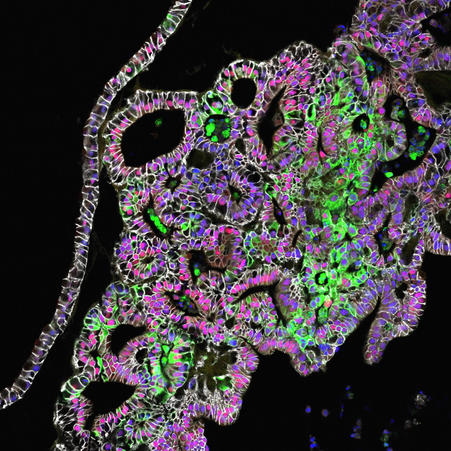
More than 20 million Canadians suffer from digestive disorders every year. Such high numbers make it necessary to study and develop new drugs to combat these diseases.  Recently, Principal investigator Jim Wells, Ph.D., director of the Pluripotent Stem Cell Facility at Cincinnati Children's and his team has generated gastric fundus organoids from human pluripotent stem cells (hPOCs)2, which will prove to be crucial to the testing of new drugs, designed to treat digestive problems.Drug testing is often carried out in mouse models, before they can be approved for clinical trials. This approach can be problematic as mice and humans differ physiologically and genetically and the drugs tested in mouse models might not always have the same effects in humans. Furthermore, once the drug is tested in an animal model then it has to go through human clinical trials, which can be limited by the number of participants. These issues can be avoided if we use organoids as models for drug testing. Organoids are miniaturized organs that show realistic microanatomy and are produced in vitro, usually from stem cells. These are genetically identical to humans and can be regenerated, hence making drug testing more convenient and reliable.

In a study published in nature, the authors generated gastric fundus organoids from hPOCs3. A key challenge the authors faced was the fact that there was no known protocol or pathway that was previously known to contribute to fundus differentiation. In this study, the authors determined that the Wnt/β-catenin pathway were essential for fundus development because knocking out β-catenin led to the expression of Pdx1, a marker found in antral epithelium of the stomach but not the fundus epithelium. Furthermore, fundus specific markers such as Irx2, Irx3, Irx5 were greatly reduced as well. Once the importance of Wnt/β-catenin was realized, the authors indirectly stimulated the Wnt/β-catenin by using CIHR. This drove the development of fundal organoids, which was then followed by the use of a variety of cytodifferentiation factors to stimulate the formation of fundal epithelium specific lineages.

This study has made key contributions to the development of a system on which drugs can be tested to determine their effects on the gastrointestinal system. Currently, our knowledge of the key pathways that regulate the differentiation of gastrointestinal progenitors into specific lineages is still incomplete. This system can shed some light onto those regulatory pathways and help us to develop even more robust models for drug testing and studying gastrointestinal diseases.

References

  1. http://www.cdhf.ca/en/statistics
  2. http://www.medicalnewstoday.com/articles/315043.php
  3. McCracken, K. W. et al. Wnt/β-catenin promotes gastric fundus specification in mice and humans. Nature 541, 182–187 (2017).


Article Summary Courtesy: Jennifer Lu and Waleed Khan

STEM CELL DERIVED CARTILAGE: A NEW ALTERNATIVE TO HIP REPLACEMENT SURGERY

Each year more than 332,000 hip replacement surgeries are performed in the United States. These hip replacement surgeries are often performed in response to osteoarthritis that weakens the hip bone overtime. The surgery involves removing parts of the hip bone and replacing them with prosthetics, which usually last up to 20 years, thus making it highly likely that a young patient will have to go through the surgery again. It is much more difficult to replace a worn out prosthetic, which can cause significant damage to the rest of the bone; therefore, there is a need for an alternative to hip replacement that can survive for longer. The solution may have come from a team of researchers from Washington University School of Medicine in St. Louis who have used stem cells to grow new cartilage.

477357450_XS

The cartilage has been grown over a synthetic scaffold that can be molded into the shape of the person’s hip. The implant can tolerate weight up to 10 times the patient’s weight and has a longer life than a traditional hip replacement prosthetic. One of the reasons for a longer life-span is because the stem cells used to generate the cartilage were transformed with a gene that allows the cartilage to produce anti-inflammatory factors when induced by a specific drug. This is important because it prevents damage to the cartilage and the scaffold when transplanted. Currently, animal trials are in progress but if all goes well, a treatment for osteoarthritis could would reach human testing in 3-5 years.

References:

http://www.medicalnewstoday.com/articles/311761.php

     Moutos, F. T. et al. Anatomically shaped tissue-engineered cartilage with tunable and inducible anticytokine delivery for biological joint resurfacing. Proc. Natl. Acad. Sci. U.S.A. 113, E4513–22 (2016).

Article Summary Courtesy: Waleed Khan

STEM CELL TECHNIQUE PREDICTS PATIENTS LIKELY TO HAVE SEVERE CHEMO REACTION

Doxorubicin is a commonly used and effective chemotherapy agent in treating a wide range of cancers such as breast cancer; however, it can cause dose-dependent cardiotoxicity in some patients. There is currently no predictory test that can be done to predict which patients will likely develop cardiotoxicity.

download

Recently, in a paper published in Nature Medicine, researchers from Stanford and Northwestern generated patient-specific human induced pluripotent stem cell-derived cardiomyocytes from skin cells in breast cancer patients who had been treated with doxorubicin and assessed for the effects of doxorubicin at various concentrations on DNA damage, calcium handling, and oxidative stress. Cardiomyocytes derived from patients experienced cardiotoxicity were found to be more sensitive to doxorubicin induced toxicity than cardiomyocytes from patients who did not experience cardiotoxicity. Sensitive cells exhibited increased reactive oxygen species production and mitochondrial dysregulation. The researchers further studied the mechanisms of action of doxorubicin. By treating the cardiomyocytes a well-known antioxidant NAC, cardiotoxicity was significantly decreased in comparison to controls, confirming that ROS-based toxicity plays an important role in the viability of doxorubicin treated cells. This model provides a platform for the discovery of new doxorubicin induced cardiotoxicity cardioprotectants.

This study has important implications for treating cancer patients. If serious cardiotoxicity can be predicted, alternative treatments or lower doses can be provided whereas high doses for better improvement can be given to patients who are likely to be resistant to related toxicity.

Summary Courtesy of Jennifer Lu 

References:

http://www.medicalnewstoday.com/articles/309302.php

Human induced pluripotent stem cell–derived cardiomyocytes recapitulate the predilection of breast cancer patients to doxorubicin-induced cardiotoxicity, Paul W Burridge et al., Nature Medicine, doi:10.1038/nm.4087, published online 18 April 2016.

ROLE OF OCT4 IN SOMATIC STEM CELLS


stem-cells

OCT4 is a transcription factor that plays a crucial role in regulating pluripotency in embryonic stem cells. It was thought that OCT4 was epigenetically silenced in adult somatic cells; however, recent evidence suggests that OCT4 might be reactivated in some somatic cell types such as tumours. A recent study published in Nature Medicine suggests that OCT4 may play an atheroprotective role when it is activated in smooth muscle cells (SMC). After vascular injury or during the development of atherosclerosis smooth muscle cells are known to undergo dedifferentiation. This process is thought to cause the repair and remodeling of blood vessels after they have been damaged.

SMCs have increasingly been shown to be involved in this process and OCT4 is shown to be reactivated in SMCs following an atherosclerosis lesion. The researchers knocked out the OCT4 from SMC cells to test for its effects on lesion pathogenesis. They saw an increase in lesion size and a decrease in lesion stability after knocking out OCT4 from SMC cells. Comparisons with Klf4-knockout studies suggest that the loss of OCT4 affects the very early phases of lesion pathogenesis, which might disturb the migration of SMCs leading to a lesion that is without a fibrous cap and is therefore destabilized. The results therefore suggest that OCT4 plays an atheroprotective role in SMC. This study is one of the first that demonstrates a functional role of OCT4 and it also suggests that OCT4 might be functionally important in other somatic cells as well.

Summary Courtesy: Waleed Khan

Reference: Cherepanova, O. et al. Activation of the pluripotency factor OCT4 in smooth muscle cells is atheroprotective. Nat Med(2016). doi:10.1038/nm.4109


SKIN CELLS CAN SWITCH BETWEEN MAINTENANCE AND HEALING STATES

The study published in Nature Cell Biology by Wellcome Trust Sanger Institute and the University of Cambridge discovered that skin cells can flip between two states, an expanding state and a balanced state. In the balanced state, the skin cells divide to produce an equal number of dividing and non-dividing cells whereas in the expanding state the skin cells divide to produce a greater number of dividing cells than non-dividing ones. According to Dr. Jones, the senior author of this study, “this research demonstrates that dividing human skin cells can switch their behavior between these two modes of maintenance or repair, challenging the longstanding view that skin renewal and healing relies on a special population of stem cells."

cell-division-telophase-250

For this study the researchers made videos of more than 3,000 skin cells dividing in culture. They showed that single cells divided in the expanding mode until they produced a multi-layered sheet of cells after which they flip into the balanced mode. They also found that these states are reversible and when a signaling protein ROCK2 kinase is inhibited, it prevents the cells in their repair mode to switch back into their maintenance mode. This finding is significant for cancer research since this could potentially prove as a mechanism by which cancers could arise.


References:
Roshan, A. et al. Human keratinocytes have two interconvertible modes of proliferation. Nat. Cell Biol. (2015). doi:10.1038/ncb3282

WORKING ON A CURE FOR AGE-RELATED BLINDNESS

12088006_519264561555803_3473906083163074172_n

   Age-related macular degeneration (AMD) is the leading cause for blindness in older people. This disease affects an area of the retina called the macula which is responsible for forming the sharpest images in the eye; hence, degeneration of this region leads to blurry vision. There are two kinds of AMD: dry AMD and wet AMD.
   Dry AMD is the most common form of AMD, and it is caused by the progressive loss of Retinal Pigment Epithelial (RPE) cells. These cells support the photoreceptors that detect light in the retina and hence are critical to vision. On the other had wet AMD is caused by the continual release of fluid from blood vessels in the retina. Wet AMD is more serious than dry AMD.

   Now the London Project to Cure Blindness is carrying out a trial in which RPE cells derived from stem cells are being transplanted in patients suffering from wet AMD via a patch. These transplants were carried last month and so far everything seems to be fine. The cells seem to be in the correct place and healthy.
   This trial will monitor 10 patients that receive the transplant over the course of 18 months. If this is successful, such technology can be used to treat AMD and hence blindness. The next report of this exciting study will come in late December. So stay tuned!


Article summary courtesy of Waleed Khan                                                                                                                                                        

"Medical News Today" Article


References:
The London Project to Cure Blindness news release, accessed 29 September 2015.
Additional source: BBC News, Stem cell trial aims to cure blindness, accessed 29 September 2015.
Additional source: Mayo Clinic, Wet macular degeneration - definition, accessed 29 September 2015.
Additional source: National Eye Institute, Facts about age-related macular degeneration, accessed 29 September 2015.


USE OF STEM CELL TECHNOLOGY GIVES NEW INSIGHT INTO THE GENETIC BASIS FOR AUTISM

12043180_516301438518782_7437684665850379112_n

   Previously many researchers have tried to understand the genetic basis for autism. This work involved comparing the genomes of autistic patients against that of people who weren’t autistic. The work hinted a few genes that might be involved in autism but a definite genetic link is yet to found.

   Recently, scientists at the Yale School of Medicine tried using a very interesting technique to tackle this problem. They isolated skin cells from autistic patients who had enlarged brains, and from their non-autistic fathers and converted them to induced pluripotent stem cells using stem cell techniques. The researchers then allowed the stem cells to differentiate into brain cells and form miniature brain like structures called organoids.

   These organoids were a good representation of the brain in its early stages of development during the first few months of gestation. By comparing the organoids from the autistic patients and those from their fathers, the researchers identified that organoids from patients with autism had a higher production of inhibitory neurons compared to those from their fathers. The researchers also identified the FOXG1 gene to be responsible for this overproduction of the inhibitory neurons. The researchers were able to correct the overproduction of inhibitory neurons by simply suppressing this gene.

   This work gives a much clearer insight into how autism might be genetically regulated and it also makes us hopeful for a cure for autism sometime in the future.


References:
Mariani, J., Coppola, G., Zhang, P., Abyzov, A., Provini, L., Tomasini, L., . . . Vaccarino, F. (2015). FOXG1-Dependent Dysregulation of GABA/Glutamate Neuron Differentiation in Autism Spectrum Disorders. Cell, 162(2), 375-390.

TINY BALLS OF HUMAN CELLS MIMIC THE BRAIN

11781883_494812804000979_264805178313998046_n

   One of the big problems with delineating neurological disorders is that we most often do not have direct access to the patient’s brain. Researchers at the Stanford University School of Medicine recently reported using iPSC’s from a patient to generate patient-specific neuronal networks is a viable option of modelling brain circuits. What this means is that, skin cells could be taken from any patient and be transformed into iPSC’s (Induced Pluripotent Stem Cells), meaning that they can now differentiate into any type of cell. Then, these can differentiate into brain cells and develop into a ball of neurons, called human cortical spheroids (hCS), of the same genetic background. This method is amazing because researchers could study the development of the neural circuitry of a specific patient and find out where things went wrong. 

   These human cortical spheroids grew to be 5mm in diameter by 2.5 months, and highly resembled a mid-fetal prenatal brain. This spherical organization were primarily driven by the cells themselves, because in dishes that made it difficult for cells to adhere to the dish itself, the cell culture began to fold themselves into spheres. Within days, these cells developed into nerve-precursors and then into fully differentiated neurons. Researchers made slices of these spheroids, much like how they would prepare mouse brain slices for analysis, and found that the architecture within these balls of neurons was strongly similar to the human cortex. As a final test, the hCS’s were put to perform functional tests, and researchers found that 80% of neurons in the spheres had the capability of firing when stimulated. Not only neurons were created from this chemical soup of iPSC’s; astrocytes, star-shaped cells that are critical for neuronal signalling, were found to be genetically identical, to surrounding neurons that they developed in concert with. This was something that has been challenging to do in the past, and now researchers will be able to generate astrocytes and neurons simultaneously and efficiently.

Article summary courtesy of Sean Ihn

References:

  1. Functional cortical neurons and astrocytes from human pluripotent stem cells in 3D culture, A. Pasca et al., Nature Methods, doi:10.1038/nmeth.3415, 25 May 2015
  2. Tiny spheres of human cells mimic the brain, researchers say

Featuring Recent Posts Wordpress Widget development by YD