TIME SPENT THROUGH THE CELL CYCLE SHOWN TO AFFECT THE FUNCTION OF STEM CELLS

1549208_492031980945728_5779433538515900391_n

   Researchers at Carl Gustav Carus Faculty of Medicine at Dresden Technical University have shown for the first time that the length of time hematopoietic stem cells spend in the G1 phase of the cell cycle determines the fitness of human blood stem cells. 
Blood is composed of many different kinds of cells, which must be continuously replaced in order to ensure survival. For this purpose we have hematopoietic (blood) stem cells in the bone marrow that have the potential to divide and differentiate into the many different kinds of cells present in blood. This process is extremely important after an infectious disease, during which a large number of blood cells are killed so they have to be replaced quickly by the stem cells in the bone marrow. This property of hematopoietic stem cells is also used in bone marrow transplants for leukemic patients so that the transplanted bone marrow can replace the diseased blood cells. However, due to resistance to the stem cells only a limited number can be transplanted for which reason it is important for us to understand the working of these stem cell.

   It is known that these stem cells are in the resting phase of the cell cycle and when more blood cells are needed they resume their cell cycle to generate more cells. However it was unknown whether the length of the cell cycle could potentially affect the functionality of the cells. To answer this researchers at Dresden Technical University use gene transfer technology to shorten the early phase of G1. They found that this significantly increased the function of the stem cells. Also the stem cells showed better function when transplanted into another mouse model. The surprise came when the researchers found that shortening the later phase of G1 had the exact opposite effect. This shows that stem cells function depends on key events that take place in the earlier and later phases of G1 and in the future scientists might be able to manipulate these events in order to achieve better stem cell function for treatments like bone marrow transplants.

Summary courtesy of Waleed Khan

References:
Blood stem cells in a rush: Velocity determines quality
NOVEL DISCOVERY TIES TOGETHER EPIGENETICS AND MALIGNANT BRAIN CANCER

   Cancer is infamous for its evasive and unrestrained proliferation, which is attributed to a smaller fraction of cancer cells called cancer stem cells. Researchers at the University of California, San Diego School of Medicine and Moores Cancer Center studied glioblastoma, a highly aggressive form of brain cancer, and noticed that genetically identical human glioblastoma cells differed in their tumorigenicity when transplanted into mice. From this, they discovered that cancer stem cell properties are defined by epigenetic changes, which are chemical modifications to the genome that cells use to control and regulate gene activity, rather than genetic changes. In this study, researchers reported that an enzyme known as Lysine-Specific Demethylase 1 (LSD1) switches off genes that are required to maintain cancer stem cell properties in glioblastoma, and that the abundance of LSD1 in the cells is the epigenetic factor that determines whether or not glioblastoma cells can propagate indefinitely as cancer stem cells.

   How does LSD1 do this exactly? Simply put, LSD1 removes methyl groups from DNA, or demethylases DNA, which turns off a number genes required for maintaining cancer stem cell properties, such as MYC, SOX2, OLIG2 and POU3F2. With these findings, Clark Chen, one of the study’s first authors, commented on how this mechanism could elucidate resistance to cancer therapy: for example, in the presence of a drug targeting MYC, glioblastomas can escape the lethal effects by shutting it off epigenetically, and turning it on once the drug is no longer present. In conclusion, this discovery leads to the possibility of creating drugs that modify LSD1 levels.

11222617_489545994527660_5718606506977386119_o

MICROTUBULE NANOTUBES MEDIATE SIGNALLING BETWEEN STEM CELLS AND NICHE IN DROSPHILA

  Stem cell niches are critical for providing resident stem cells with signals that specify their identity, and their signals only act over short distances so that they reach the progenitor cells only, not their differentiated progeny. However, the molecular mechanisms of this niche signalling were poorly understood. This recent study used Drosphila male germ line stem cells (from their testes) to identify and characterize microtubule (MT) -based nanotubes that extend from these cells into the hub, the central region of the niche. It also reported a finding that MT-nanotubes were specific only to germline stem cells. The hub secretes at least two ligands, one being a bone morphogenetic protein (BMP) ligand called Decapentaplegic (Dpp), which regulates the maintenance of germline stem cells.

  Research has found that the interaction between these secreted Dpps, and BMP receptors called Thickveins (Thv), which are expressed in germ cells, is necessary for the formation of these MT-nanotubes. Also, the disruption of MT-nanotubes jeopardizes the Dpp signalling within germline stem cells, leading to the germline stem cell loss. Possible reasons why such a structure was not discovered before include the fact that they are sensitive to fixation, not always associated with the centrosome, and lacking acetylated microtubules. In sum, the ligand-receptor interaction (Dpp-Thv, respectively) promote MT-nanotube growth, which further enhances the signalling within stem cells in a niche, and leads spatially oriented self-renewal.


Article summary courtesy of Sean Ihn

Reference:
Nanotubes mediate niche-stem-cell signalling in the Drosophila testis, Mayu Inaba et al., Nature, doi:10.1038/nature14602, 1 July 2015

COLORECTAL CANCER SPREAD MAY BE HALTED BY TARGETING DORMANT CELLS

Colorectal cancer spread is among the leading causes of cancer related deaths. Usually, people die from secondary tumors that develop from colorectal cancer rather than the primary tumor itself. By the time the tumor is detected the cancer has already spread, making it extremely difficult to treat. The cancer has poor diagnosis because the cancer cells can stay dormant for long periods of time until some signal causes them to activate and start growing and dividing rapidly.
Now a study published in Stem Cell Reports suggests a possible way for targeting the cancer cells while they are dormant. According to past research, there is a stem cell in the gut that is identifiable by its unique Lgr5 receptor, which is thought be responsible to activating the tumor cells. The stem cells are called crypt base stem cells, which allow the passage of a ‘WNT’ signal that can activate the tumor cell through a cell surface receptor called Frizzled. However, until now it was not known which type of Frizzled receptor led to this activation. There are 10 different types of Frizzled receptors, and through this research the scientists have been able to demonstrate that Frizzled7 is responsible for terminating the dormancy of the tumor cell. This provides us with a novel target for attacking colorectal cancer cell even in their dormancy, which can significantly improve colorectal cancer treatments.

Article summary courtesy by Waleed Khan

Article: Medical News Today

EPIGENETIC CHANGES THAT CONTROL ENDOTHELIAL CELL DEVELOPMENT

Researchers at the University of Illinois have recently discovered epigenetic changes that might be involved in endothelial cell development. Epigenetic changes are non-genetic changes that can affect the way genes expressed. One key way epigenetic changes occur is through modification of histones, proteins around which DNA is bound. The modification involved in this case was the demethylation of histones that appears to be crucial for the conversion of stem cells into endothelial cells.
This study identified two enzymes that were necessary for the regulation of certain genes needed for the transformation of embryonic stem cells into endothelial cells. These two enzymes, KDM4A and KDM4C, are histone demethylases and alter the expression of these genes with epigenetic modifications. The researchers initially observed high levels of these two demethylases in mice during the transformation from embryonic stem cells to endothelial cells, but further corroborated this finding in zebrafish, by knocking down these enzymes in embryos and observing their inability to form blood vessels.
The researchers believe that the demethylation of histones affects a number of promoters for genes involved in endothelial cell development. This study will allow researcher to gain more knowledge about this process that researchers can then use to develop endothelial cells for uses such as tissue repair.

Article summary courtesy by Waleed Khan and Sean Ihn

Article: ScienceDaily

Reference: 
Liangtang Wu, Kishore K. Wary, Sergei Revskoy, Xiaopei Gao, Kitman Tsang, Yulia A. Komarova, Jalees Rehman, Asrar B. Malik. Histone Demethylases KDM4A and KDM4C Regulate Differentiation of Embryonic Stem Cells to Endothelial Cells. Stem Cell Reports, 2015; DOI: 10.1016/j.stemcr.2015.05.016

FULL LIMB GENERATION

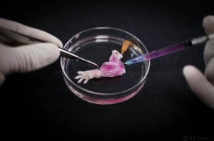
Despite improving prosthetic technology, people who have lost a limb still face limitations. One alternative to prosthetic limbs would be limb transplant; however, this can lead to a prolonged high risk of immune rejection, which can make life even harder. Now researchers at Massachusetts General Hospital in Boston have been able to circumnavigate this issue by growing limbs by using an individual’s own cells rather than a transplant from a donor. Limbs involve a complex system of muscles, bones, vessels and nerves and in order for them to develop and grow properly; they need a supporting structure called a matrix.

Full Limb Generation

In order to establish this, the researchers stripped away cells from the forelimbs of dead rats, but preserved the vascular and neural matrix. The researchers then repopulated these with progenitor cells and allowed them to grow and develop in a bioreactor. The limb was removed from the reactor after two weeks and tested for functionality. Electrical stimulation of the muscles in the limb caused the muscles to contract appropriately and once the limb was transplanted in another rat blood started to immediately flow into the new limb. This is a great step towards artificially growing limbs and using them for transplants. The researchers are now focusing on trying to ensure that neural connections are correctly made between the nerves of the limb and of the recipient organism.

Article summary courtesy by  Waleed Khan

References:
Engineered composite tissue as a bioartificial limb graft, Harald C. Ott et al., Biomaterials, doi:10.1016/j.biomaterials.2015.04.051, published online 2 June 2015

NOVEL DIABETIC NEUROPATHY TREATMENT USING STEM CELLS

Diabetic neuropathy is the degeneration and the demyelination of nerves, particular in the lower regions of the body, due to high blood sugar levels. Current treatment options available only mitigate the discomfort and distress of the patients, and strictly regulate blood sugar levels. This article that was published today in GEN (Genetic Engineering News) outlines the discovery from researchers from South Korea and the U.S. in regards to the novel therapeutic potential in using mesenchymal stem cells (MSC) from bone marrow to treat diabetic neuropathy. The injection of bone marrow MSCs into rats displayed successful migration of the cells to the diabetic nerves, as well as angiogenesis, re-myelination, and axonal regeneration. This study showcases the promising potential in cell therapy as a possible therapeutic option for a disease that has previously rendered diabetic patients hopeless.

Article summary courtesy by Sean Ihn

Article: Genetic Engineering News (GEN)

DEATH OF A WHITE BLOOD CELL FILMED FOR THE FIRST TIME

Researchers from La Trobe University used time-lapse microscopy to record the death of a monocyte, a kind of white blood cell. Previously, researchers believed that white blood cells randomly broke apart during death; however, this study showed that as the cell died it produced structures that are being called ‘beads’.

The cell produced a long bead like protrusions that look like beads on a string. This then breaks into individual beads. Scientists believe that not only do these beads make it easier to clear up the cell debris but they may also contain molecules that alert the other cells of the immune system of a pathogen that has infected the body. The researchers also think that certain viruses might hide in these beads and then use them to spread in the body. Another interesting thing that they found was that beads’ formation was blocked by certain antidepressants whereas a certain antibiotic can promote their formation.

Summary courtesy by  Waleed Khan

Reference: Nature Communications PDF


STEM CELL DEVELOPMENT IS CONTROLLED BY THE MITOCHONDRIA

Researchers at the NYU Langone Medical Center recently published a study in Nature Cell Biology in which they investigate the role of ATP synthase in the development of stem cells that develop into eggs and sperms in fruit flies. ATP synthase is an enzyme present in the mitochondria that produces ATP during the process of cellular respiration to power the chemical reactions in the cell. The researchers discovered that blocking the action of ATP synthase prevented the development of the stem cells responsible for germ cell development. Since ATP synthase is involved in the process of oxidative phosphorylation, the researchers wanted to determine whether ATP synthase or the process of oxidative phosphorylation were responsible for stem cell development.

Mitochondrial Control in Stem Cell Growth

They tested this by blocking other enzymes involved in the pathway, but they found that it did not stop stem cell development. As a result the researchers were able to conclude that ATP synthase played a role in this process. In fact disrupting any of the 13 proteins involved in ATP synthase disrupted stem cell development. The research also shows that ATP synthase could be involved in the development of cristae, folds in the inner membrane of mitochondrion, as the stem cell divides. Previous studies have shown that often mitochondria with faulty cristae have faulty ATP synthase; however, this study was able to provide a more concrete proof of this. The researchers are now interested in investigating the exact mechanism by which ATP synthase controls cristae development in the mitochondria.

Article summary courtesy by Waleed Khan

Article: Medical News Today

Reference: Nature

A SINGLE STEM CELL INJECTION SLOWS AMD VISION LOSS

Recently, scientists Cedars-Sinai Medical Center in Los Angeles, CA, have shown that they can slow down vision loss due to AMD by a single stem cell injection. AMD (Age-related macular degeneration) is a degenerative disease that occurs due to the deterioration of the macula, the central part of the retina that is responsible for sensing light. People who suffer from AMD gradually lose their vision completely and there is no known cure for this condition.
But scientists at Cedars-Sinai Medical Center have been able to significantly slow this process in AMD rats. The created induced pluripotent stem cells (iPSCs) from epithelial cells and the converted these iPSCs into induced neural progenitor stem cells (iNPSCs). On injecting these iNPSCs cells into AMD rats , the AMD progression slowed by 130 days which is equivalent of 16 years in humans. This study shows the amazing promise of stem cells in treatments for AMD.
The team now aims to carry out preclinical trials on animals to test the efficiency and safety of stem cells injection before clinical trails can begin on people who are suffering from later stages of AMD.

Article summary courtesy by Waleed Khan

Article: Medical News Today

References: 
Human iPSC-derived neural progenitors preserve vision in an AMD-like model, Yuchun Tsai, et al., Stem Cells, doi:10.1002/stem.2032, published online 13 April 2015, abstract.

Featuring Recent Posts Wordpress Widget development by YD