AMNIOTIC STEM CELLS IN COMBINATION WITH HYDROGEL THERAPY BOOST BLOOD VESSEL GROWTH IN MICE

A team led by Jeffrey G. Jacot, associate professor of bioengineering at Rice University recently demonstrated that amniotic stem cells have the potential to be used as a therapeutic for heart diseases. Amniotic stem cells are cells that are taken from the amniotic fluid and have the ability to give rise to a wide variety of cells, including endothelial cells that can form blood vessels.
The team used hydrogels seeded with amniotic stem cells to see if it can promote the formation of new blood vessels. Hydrogel 3D scaffolds are widely used in medicine to deliver drugs and one form of it that contains fibrin can promote angiogenesis. The team modified the hydrogel with polyethylene glycol that makes it firm and long lasting.

Hydrogel Therapy with Amniotic Stem Cells

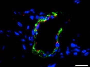
The team then injected mice with either the hydrogel or with the hydrogel seeded with the stem cells. Growth factors were also added to promote cellular differentiation. The team found that the hydrogel seeded with the stem cells led to greater growth of blood vessels. This is significant since these fibrin hydrogels in combination with amniotic stem cells can be used to treat heart diseases. At the same time, if the stem cells are taken from the amniotic fluid of the infant, which has to receive treatment, it will reduce the chances of immune rejection. Right now, the team is working on developing biocompatible patches that can be used to treat infants with defects in their heart.

Article summary courtesy by Waleed Khan

Article: Medical News Today

Reference:

In situ vascularization of injectable fibrin/poly(ethylene glycol) hydrogels by human amniotic fluid-derived stem cells, Omar M. Benavides et al., Journal of Biomedical Materials Research Part A, doi:10.1002/jbm.a.35402, published online 9 February 2015

THE GENE FOR THE MOST AGGRESSIVE FORM OF BREAST CANCER HAS BEEN IDENTIFIED

A group of researchers have determined that the ‘Inhibitor of Differentiation 4’ (ID4) gene is not only associated with a highly aggressive triple negative breast cancer but also controls it. Triple breast cancer is type of breast cancer that lacks estrogen, progesterone and HER2 receptors. These receptors are used by many treatments to target cancer cells. A lack of these receptors also leads to poorer prognosis. Those with triple breast cancer either die quickly i.e. in 3-5 years or they stay alive for much longer. The apparent difference can be attributed to whether the cancer developed from specialized cells or stem cells, and the latter leads to the more aggressive one.

Breast Cancer Gene

The researchers blocked the expression of ID4 and noticed that it led to the activation of other genes that lead to specialization. It also led to the expression of estrogen and progesterone receptors that would potentially improve the prognosis of triple negative breast cancer. However, it is still not clear whether the receptor is actually an estrogen receptor or is it a merely a similar structure. The team is now planning to work with an expert on estrogen receptor function to determine the exact biochemical roles that ID4 plays in the development of cancer.

Article summary courtesy by Waleed Khan

Article: Medical News Today

References:

 ID4 controls mammary stem cells and marks breast cancers with a stem cell-like phenotype, Alex Swarbrick, et al., Nat. Commun., doi:10.1038/ncomms7548, published online 27 March 2015, abstract.


NEW HOPES FOR THE ‘BUBBLE BOY DISEASE’

Severe combined immunodeficiency disease (SCID) is a group of rare genetic disorders that prevent the bone marrow from making important immune cells such as the natural killer (NK) cells and T cells. The lack of NK and T cells severely disrupts the immune system and renders the person defenseless even to the mildest of infections. Children born with SCID are often covered in a bubble bag, hence the name ‘bubble boy’, in order to maintain a sterile environment that protects them from pathogens.
Previously, gene therapy was considered a promising technique to treat SCID but when viruses were used to deliver correct genes to the bone marrow, often leukemia developed and so scientists had to resort to other forms of gene therapy that only benefitted those with mild SCID.
Now a research team from Salk Institute of Biological Studies has managed to generate NK cells from genetically edited stem cells of an SCID patient.

Bubble Boy Disease

The process targets a very specific form of SCID known as X-linked severe combined immunodeficiency disease (X-SCID) by obtaining cells from the patient and then converting them to induced pluripotent stem cells (iPSCs). The iPSCs are then genetically modified at specific points using the gene editing technology known as TALEN, that involves the use of enzymes that can cut out individual nucleotides at specific points to correct the X-SCID causing faulty genes. These cells are then treated to produce NK cells. This is a significant breakthrough, as this process will ensure immune compatibility as well as an unlimited supply of NK cells (since stem cells can divide indefinitely).
Despite its promise, this process is still in its early stages and scientists still need to figure out how to generate T cells that are also scarce in SCID patients. The research team has been able to generate a precursor of a T cell but generating a mature T cell from the gene-edited iPSCs cells is yet to be done.

Article: Medical News Today

Research Paper: Cell Stem Cell

Article Summary Courtesy of Waleed Khan

HEART ON A CHIP BRINGS US CLOSER TO BETTER DRUG TESTING

Most drugs that are developed to target the heart are often tested on animal models. This method is not very efficient due to fundamental differences in the biology of humans and other living organisms such as the ion channels that conduct electrical impulses in the heart, which most of these drugs target. This limitation often delays the release of the drug and often incurs huge costs; however, the ‘heart-on-a-chip’ model developed by Professor Haley and his colleagues at UC Berkeley might just be the answer to this problem.

Heart on a chip
The heart-on-a-chip comprises of heart cells created from induced pluripotent stem cells that are arranged in a 3D geometry. Microfluidic channels on either side of the cellular area mimic blood vessels and can be used to deliver drugs the cell. This ensures that the cells are exposed to the nutrients and drugs in a way similar to that inside the body. The heart-on-a-chip was tested with four drugs and was able to replicate the effect of the drugs would have had on the heart inside the body, which was done by measuring the pulse rate of the cells.
The team is now planning to investigate whether this method of drug testing will work in multi-organ interactions. If this technology advances successfully it could possibly replace the use of animal models of drug testing in hopes of more reliable results.

Article: Medical News Today 

Research Paper: Nature

Article summary courtesy by Waleed Khan

STEM CELLS BRING US ONE STEP CLOSER TO REPAIRING CARTILAGE

Cartilage is a connective tissue found in joints, the ears, nose etc. It plays the essential function of holding bones together and keeping various passageways open in the body, such as the trachea. However, as people grow old the cartilage starts to wear, especially in joints that have experienced constant stress, leading to a degenerative disease known as Osteoarthritis.

Cartilage Repair
Researchers at the University of Manchester have recently reported that they successfully produced cartilage in rats from embryonic stem cells. The researchers generated chondroprogenitors, precursor to cartilage cells, from human embryonic stem cells and then implanted the cells in the knee joints of rats that had damaged cartilage. The cartilage was partially repaired after only four weeks and attained a similar appearance to normal cartilage by week twelve. Not only did the cartilage develop normally, but also there was no abnormal tumor growth, which is a common occurrence. This is promising research since it can potentially lead to new ways of repairing damaged cartilage. This experiment on rats will eventually pave the way for trails in humans that are still some time away in the future.

Article: Medical News Today


Article summary courtesy of Waleed Khan

GENERATING ARTIFICIAL HUMAN PRIMORDIAL GERM CELLS FROM PLURIPOTENT STEM CELLS

Scientists from Israel and the UK have for the first time generated artificial human Primordial Germ cells (PGCs) from induced Pluripotent stem cells.

PGCs are precursors cells that eventually develop into sperm and egg cells in the testes and the ovaries respectively. Previously, in 2012, Mitinori Saitou of Kyoto University in Japan and his collaborators first created artificial PGCs from pluripotent stem cells derived from mice skin cells. Although, these cells cannot be cultured into mature gamete cells in vitro but the researchers suspect that they can generate sperm or egg cells if transplanted in the gonads. Scientists have since then been trying use the same method to create artificial PGCs from human induced pluripotent stem cells; however, the process is more complicated than it seems. This is because mouse and human stem cells are different from each other.

Artificial Primordial Germ Cells

It’s only recently that a research team led by Azim Surani of the University of Cambridge, UK, and Jacob Hanna of the Weizmann Institute of Science in Rehovot, Israel were able to replicate the in vitro part of this process in human stem cells. The cells that they generated have all the markers of Primordial Germ cells. This development is huge because it may provide a way for sterile individuals to have children. However, this technology is far from being implemented. The next step is to implant these cells in the testes and ovaries of mice to see if they can develop into mature gamete cells.

Article: ScienceDaily

Research Paper: Nature

~ Article summary courtesy of Waleed Khan


COMBATING CANCER STEM CELLS WITH GRAPHENE OXIDE

Researchers in the University of Manchester have recently demonstrated that graphene oxide has anti-cancer properties. Cancer is a devastating condition in which cells undergo uncontrolled mitotic divisions and damage the body. Conventional treatments for cancer usually involve chemotherapy and radiotherapy that although are able to kill the bulk of cancer cells, but do not always kill cancer stem cells, precursor cells that can renew and develop into cancer. These stem cells are difficult to identify and are highly resistant to treatment; and as a result, if left untreated in the body they can often cause the cancer to spread throughout the body by a process known as metastasis.

Graphene Oxide Therapy

To combat these cells researchers were working with a graphene oxide, a modified version of graphene. Graphene is already considered as a potential material for drug delivery because it can effectively bind to cell surfaces, but in this case graphene oxide itself was able to act as an anticancer drug. It selectively targeted these cancer stem cells and prevented them from forming tumor-spheres. Graphene oxide was effective against six different types of cancers and is now being considered as a potential therapeutic for cancers. However, for this to happen a lot of research still needs to be done and it may take a while before human trials can begin.

Article: Medical News Today

Research Paper: Oncotarget

~ Article summary courtesy of Waleed Khan


Featuring Recent Posts Wordpress Widget development by YD